Neuroscience

News

Introducing NYOX – One Platform for Field Monitoring

Discover NYOX – Skope’s new platform set to transform field monitoring practices. NYOX delivers unmatched connectivity, ease of use, and productivity and empowers our users to elevate their workflows and achieve new levels of efficiency.

Play Video

Resources

New Blog Post on How to Represent Magnetic Fields for MR Imaging

Explore the hidden complexities of magnetic field encoding in MRI, from imperfect gradients to spherical harmonics, and learn how new techniques are enhancing image clarity and diagnostic precision.

News

NEUROQUANT Project Awarded Prestigious Grant

The project NEUROQUANT – Neurovascular MR coil powered by quantitative neuroimaging technologies for Alzheimer’s disease diagnosis, prognosis, and therapy monitoring, has been awarded a prestigious grant through a joint initiative from Innovate UK and Innosuisse – Swiss Innovation Agency. Skope is one of the project partners.

Resources

Connectome 2.0 & Initial Experience with the Skope System

Learn how advances in MRI physics and technology, driven by increasing magnetic field strengths and gradient power, are revolutionizing diffusion MRI to uncover the intricate pathophysiology of diseases.

Play Video

Resources

NeuroCam™ 3T & 7T DICOM Viewer

Our new DICOM Viewer now includes images acquired with the NeuroCam 3T Excellence and the NeuroCam 7T Standard. Take a look at the latest images!

T2-Weighted Imaging: Hippocampus
Susceptibility-Weighted Imaging

Focus on Neuroscience

Image quality is at the heart of neuroimaging studies. Images which do not accurately reflect the anatomy and physiology of the subject do not contribute meaningful data to a study. Image artifacts obscure the data needed for the study, increasing the number of subjects, cost, and length of the study. Accounting for these artifacts post-acquisition can mean resorting to complex post-processing steps which may only partially remove the artifact - possibly without improving the research value of the image.

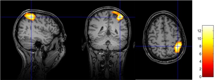
Skope develops advanced imaging solutions tailored to meet the critical needs of neuroscientists in their quest to unravel the brain structures and function, by providing high-quality, reproducible imaging for precise analysis and insight. These solutions are essential for functional MRI (fMRI), diffusion/DTI studies, and other scan advanced imaging protocols ensuring reliable data with minimal workflow disruption and mitigating scanner performance issues. 

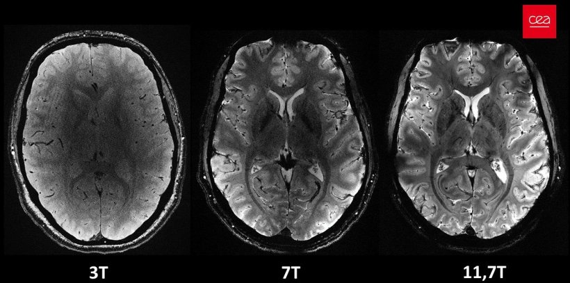
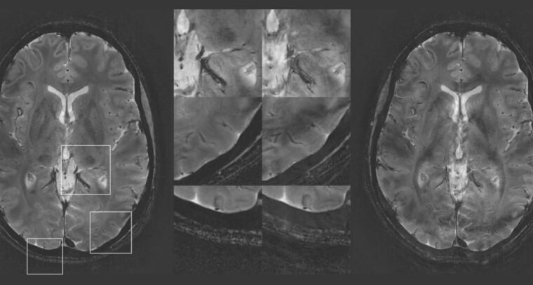
Skope’s technology accurately measures scanner performance, capturing deviations in image encoding caused by real-world scanner and subject sources. These field measurements are then used to produce images with consistent geometry, fewer artifacts, and improved signal-to-noise ratio (SNR). Images generated with Skope tools, because scanner and subject induced sources are greatly reduced, better reflect the underlying physiology being imaged.

Utilizing Skope tools has allowed neuroimaging researchers to both better utilize well-proven imaging techniques such as the Human Connectome Project protocols and pursue state-of the art techniques, such as non-linear diffusion encoding. One of Skope’s critical innovation and development directions is to enable crucial progress in understanding, diagnosing, and treating neurodegenerative diseases such as multiple sclerosis (MS) and Alzheimer's, helping to advance research that leads to better outcomes for patients.

Products Overview

NeuroCam™ 7T

MR Head Coil for Parallel Transmission with 64 Independent Receive Elements and 16 Field Probes for High Image Quality and Stability at 7T.

Relevant resources

References

I want to thank the Skope team for the fantastic collaboration throughout our project. I truly appreciated the ‘Swiss precision’ in defining the project and reliably delivering on promises made. Additionally, having the team show up on-site really gave the project an extra push, helping us meet critical milestones.

Professor Nikolaus Weiskopf, PhD

MPI for Human Cognitive and Brain Sciences

Being able to directly visualize the gradient moments made it easy to identify the error and fix the calculation in the pulse sequence.

Lars Mueller, PhD

University of Leeds

Oscillating gradient spin-echo (OGSE) diffusion acquisitions offer unique microstructural insight but suffer limited sensitivity due to the characteristically long TEs. This shortcoming can be mitigated by combining OGSE with spiral readouts…

Eric S. Michael

Institute for Biomedical Engineering, ETH Zurich

Having access to the raw trajectory data from the Skope system and libraries with which to read them allowed us to successfully develop our model-based reconstruction algorithm and generate high-fidelity images.

Paul Dubovan

Department of Medical Biophysics at Western University

Our Clip-on Camera has been invaluable to monitor field stability and fluctuations in MR thermometry. If thermal measurements are to remain non-invasive in vivo, it is impossible to obtain definite proof without in situ measurements.

Nicolas Boulant, PhD

NeuroSpin/CEA

Sign up

By submitting your email address, you agree on receiving news around and about Skope and to the Terms & Conditions and Privacy Policy.