Related Applications: Diffusion Imaging, fMRI
NeuroCamTM 7T with its 64 highly sensitive receive elements provides new opportunities for neuroimaging at 7 Tesla. The tight-fitting geometry ensures optimal coil-head coupling, maximizing sensitivity and minimizing signal loss. Additionally, it features an 8 or 16 channel transmit coil, enabling parallel transmission to achieve a homogeneous spin excitation. By elevating SNR and enabling finer spatial resolution, NeuroCamTM 7T captures subtle anatomical details and facilitates the detection of intricate brain activity.
The transmit array and the anterior part of the receiver array can be shifted toward the service end of the magnet for easy subject positioning. The top part of the receiver has indentations on both sides to easily slide it over the subject.
The detachable front-view mirror can be placed on top of the transmit coil to allow the subject to see the magnet room on the patient-side of the bore. The position on the coil can be adjusted via a sliding mechanism.
The compact design and the light weight (14 kg) of the coil assembly facilitates the handling of the coil by the operator.
The tight-fitted receiver array, which comprises 64 independent elements, provides high SNR for neuroimaging applications.
The increase in SNR is particularly evident within cortical regions when compared to 7T head coils currently available on the market.
The NeuroCamTM 7T also supports higher acceleration factors than the default 7T head coil due to its higher encoding power.
Susceptibility Weighted Imaging (SWI) at ultra-high field strengths, such as 7T, offers exceptional detail and contrast, making it ideal for visualizing intricate structures. SWI is particularly useful for detecting microbleeds and hemorrhages, visualizing veins and venous structures, identifying calcifications, and assessing iron deposition. The NeuroCamTM 7T further enhances these advantages by providing excellent image quality with fine detail.
The MPRAGE sequence provides great anatomical detail and contrast, making it ideal for visualizing fine structures in the brain and spinal cord. The shown sagittal image demonstrates the extensive coverage down to C6, showcasing the NeuroCamTM 7T's capability to capture detailed images of both the brain and upper cervical spine. The Universal pTX pulses enable uniform excitation and improved image quality across diverse populations without the need for individual patient B1 maps.
The transverse Turbo Spin Echo (TSE) images acquired in Circular Polarized (CP) mode demonstrate excellent image quality, even without the use of advanced RF shimming techniques. Despite being Specific Absorption Rate (SAR) intensive, the efficiency of the coil ensures that it can still produce excellent images at ultra-high field strength.
The 8 or 16 independent transmit elements of NeuroCamTM 7T allow to homogenize the transmit field via parallel transmission. The coil version with 16 transmit elements does not comprise a front-view mirror due to the smaller size of the transmit elements, but it includes the same rear-view mirror for visual stimulation as the 8-channel version of the coil.
The coronal TSE image demonstrates excellent left-right symmetry, achieved through the use of parallel transmission to homogenize the transmit field. The image quality is excellent, showcasing fine anatomical details. Notably, on the patient's right side, the cochlea is clearly visible.
MP2RAGE is used for obtaining high-quality T1-weighted images, providing superior contrast between white matter and grey matter. This sequence is particularly useful for visualizing sub-thalamic nuclei, deep grey matter structures, and cortical grey matter, as well as for applications such as segmentation and voxel-based morphometry. A patient-specific RF shim was used to further enhance image quality and uniformity.
FLAIR imaging at 7 Tesla (7T) presents unique challenges, primarily due to issues with magnetic field homogeneity and specific absorption rate (SAR) limitations. Enhancing image homogeneity can be achieved through efficient coil design and the use of parallel transmission techniques as demonstrated below.
A detachable rear-view mirror can be attached to the enclosure of the receiver array for visual stimulation.
The enclosure of the receiver array is designed to minimally obstruct the visual field.
The NeuroCamTM 7T is ideally suited for functional MRI (fMRI) applications. Its high cortical signal-to-noise ratio (SNR) and low g-factor penalty enable the acquisition of high-quality images with significantly reduced artifact levels compared to the standard 32-channel head coil.
The NeuroCamTM 7T is available with a fully integrated array of 16 fluorine field probes for concurrent measurement of the encoding fields. The probes are located in the enclosure of the receiver array and distributed around the imaging volume to provide optimal conditioning for the spatial expansion of the spherical harmonics basis fields. The housing of the frontend electronics for the field probe array is attached to the back of the coil, where it does not obstruct the visual field of the rear-view mirror.
The field probe hardware of the NeuroCamTM 7T can be easily connected to NYOX, the field monitoring system, using the provided multiconnector cable. This cable is detachable on both ends. If the planned scan session does not include field monitoring, the cable does not need to be connected.
Irrespective of the used encoding scheme (e.g. EPI, spiral, radial), the fully integrated field probe array allows to acquire images with enhanced stability and reproducibility.
Concurrent field monitoring enables the use of optimized sampling patterns for data acquisition. The technology facilitates applications that are particularly challenging at 7T, such as diffusion-weighted imaging. By continuously tracking the dynamic magnetic field variations, field monitoring ensures higher accuracy and consistency in the acquired data. This leads to improved image quality and resolution, making it possible to visualize fine structural details and subtle pathological changes that are critical for advanced neuroimaging studies.
By monitoring the magnetic field, real-time adjustments can be made to compensate for fluctuations caused by breathing or other factors, ensuring a more stable and uniform field throughout the imaging procedure. Alternatively, corrections can be applied during image reconstruction by incorporating the measured field data into the process. The figure below illustrates frequency fluctuations caused by a breathing subject in a 7T MR scanner, as measured by four field probes positioned at different locations within the scanner bore (Courtesy of DZNE Bonn, Germany).
The NeuroCamTM 7T users benefit from comprehensive technical documentation, providing them with substantial flexibility and the capability to conduct additional RF simulations. This documentation encompasses details on coil design, CAD models, bench and scanner test results, MR compatibility information, simulation reports, and more.
Image reconstruction that takes the actual encoding fields into account.
Enhance your existing coil with concurrent field monitoring and simply subtract dynamic field errors.
Everything you need to know about your encoding fields, measured with the latest field-probe technology.
Data management application that improves field monitoring workflow by automating data transfer and data combination.
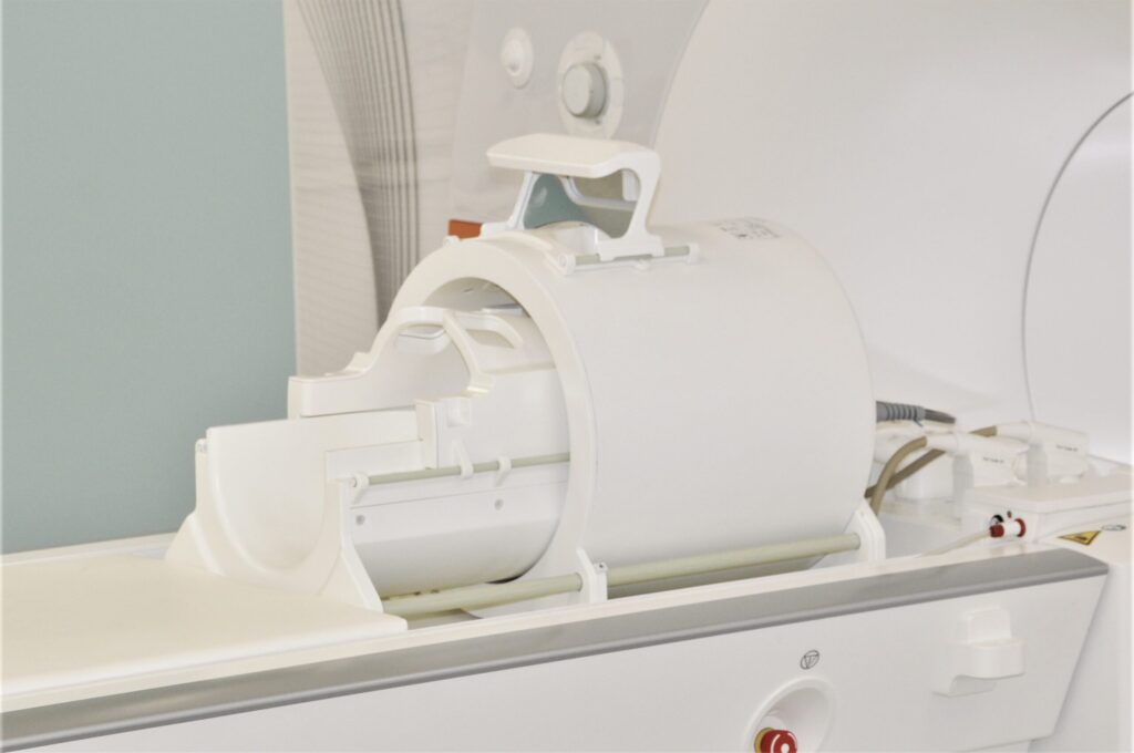
Cutting edge head coil with field monitoring, developed for neuroscientists who want to increase image quality.