Resources
New Knowledge Hub for Users
We’ve just published Skope Wiki – an extensive knowledge hub to help our users thrive. While we continue to add new articles, all users can already benefit from a wide range of general and scanner-specific topics. From getting started tips to advanced resources for maximizing the impact of Skope’s products in your lab, it’s all there! First-time registration is required.

News
Skope Achieves ISO 14001
Skope has successfully achieved the globally recognized ISO 14001 certification for its Integrated Management System (IMS). The certification is a significant milestone, formally confirming the company’s commitment to reducing its environmental footprint and operating sustainably.

News
Introducing NYOX – One Platform for Field Monitoring
Discover NYOX – Skope’s new platform set to transform field monitoring practices. NYOX delivers unmatched connectivity, ease of use, and productivity and empowers our users to elevate their workflows and achieve new levels of efficiency.
Resources
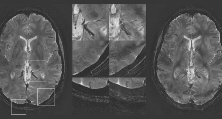
NeuroCam™ 3T & 7T DICOM Viewer
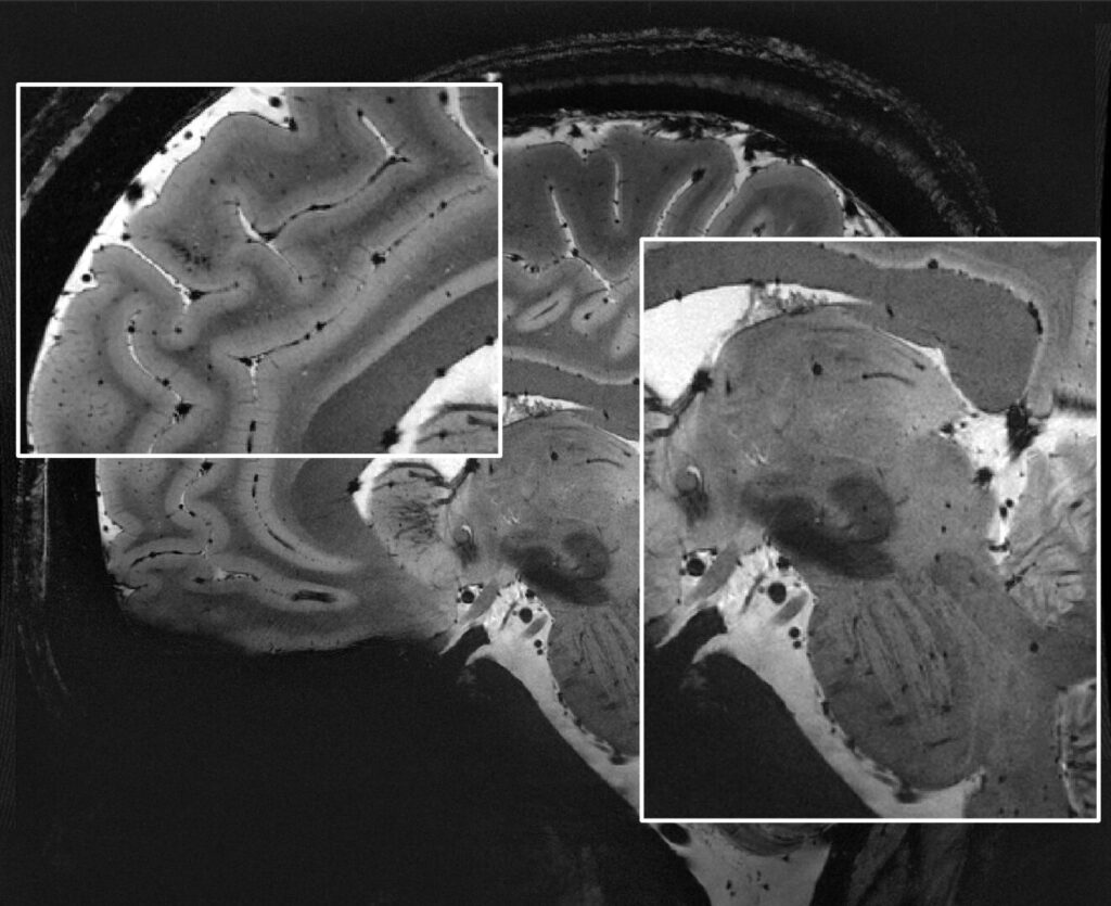
Our new DICOM Viewer now includes images acquired with the NeuroCam 3T Excellence and the NeuroCam 7T Standard. Take a look at the latest images!

Your Partner in MR Imaging
We develop innovative solutions in collaboration with the world's best MR technology scientists and engineers to deliver the highest image quality and reproducibility at the required speed for medical research and clinical applications.
Our products and solutions are vendor-agnostic.
Products Overview
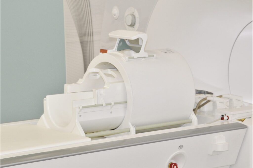
NeuroCam™ 7T
MR Head Coil for Parallel Transmission with 64 Independent Receive Elements and 16 Field Probes for High Image Quality and Stability at 7T.
NeuroCam™ 3T
Cutting edge head coil with field monitoring, developed for neuroscientists who want to increase image quality.

Clip-on Camera “Cranberry” Edition
The Clip-on Camera “Cranberry” Edition comes fully configured to operate in your 7T scanner, which improves workflow for configuration and integration. Based on our well-established Clip-on Camera, “Cranberry” delivers our neuroimaging solution right at the doorstep of your 7T scanner.

Clip-on Camera™
Enhance your existing coil with concurrent field monitoring and simply subtract dynamic field errors.

Dynamic Field Camera™
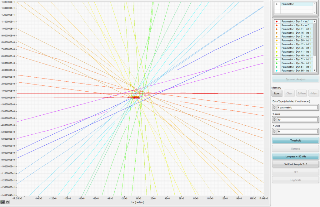
Everything you need to know about your encoding fields, measured with the latest field-probe technology.

skope™-i
Image reconstruction that takes the actual encoding fields into account.
skope™-dm
Data management application that improves field monitoring workflow by automating data transfer and data combination.

References

I want to thank the Skope team for the fantastic collaboration throughout our project. I truly appreciated the ‘Swiss precision’ in defining the project and reliably delivering on promises made. Additionally, having the team show up on-site really gave the project an extra push, helping us meet critical milestones.
Professor Nikolaus Weiskopf, PhD
MPI for Human Cognitive and Brain Sciences

Being able to directly visualize the gradient moments made it easy to identify the error and fix the calculation in the pulse sequence.
Lars Mueller, PhD
University of Leeds

Oscillating gradient spin-echo (OGSE) diffusion acquisitions offer unique microstructural insight but suffer limited sensitivity due to the characteristically long TEs. This shortcoming can be mitigated by combining OGSE with spiral readouts…
Eric S. Michael
Institute for Biomedical Engineering, ETH Zurich

Having access to the raw trajectory data from the Skope system and libraries with which to read them allowed us to successfully develop our model-based reconstruction algorithm and generate high-fidelity images.
Paul Dubovan
Department of Medical Biophysics at Western University

Fast, non-Cartesian imaging is simpler than ever since we rely on the Dynamic Field Camera for system characterization and trajectory measurement. Real-time field monitoring with a Clip-on Camera has become a cornerstone of our 7T program.
Prof. Klaas P. Prüssmann, PhD
Institute for Biomedical Engineering, ETH Zurich

At the UMC Utrecht we have built up quite some experience with the 16-channel Skope system for monitoring B0 field fluctuations in the human body at high field. We strive to stabilize sensitive MRI and MRS/MRSI acquisitions by correcting the acquisition and reconstruction techniques for B0 instabilities caused by (involuntary) patient movement.
Prof. Dennis Klomp, PhD
UMC Utrecht

We are end-users of this technology who expect ease of use. Our installation was a seamless process during which both sales team and the MR experts at Skope were a pleasure to work with.
Zoltan Nagy, PhD
Laboratory for Social and Neural Systems Research - SNS Lab, University of Zurich

The Skope staff have always been really helpful in sorting out any issues that I faced as my project developed over the past 5 years. The field camera is a very friendly solution that gives room to personalize data acquisition thanks to the MatLab-package scanner interface.
Laura Bortolotti, PhD
University of Nottingham

Our Clip-on Camera has been invaluable to monitor field stability and fluctuations in MR thermometry. If thermal measurements are to remain non-invasive in vivo, it is impossible to obtain definite proof without in situ measurements.
Nicolas Boulant, PhD
NeuroSpin/CEA
Applications Overview
Neuroscience Workflow
Solution for neuroscientists by neuroscientists.
- Download Data Sheet
MR Methods Development
Developing new MR hardware and methods requires dedication, innovation, and a lot of debugging. Field monitoring gives you a direct view of your scanner's performance to reduce guesswork and frustration in finding those bugs.
Diffusion Imaging
Improve image reproducibility and fidelity by measuring and correcting images for diffusion encoding field distortions.

Ultra-High-Field Imaging
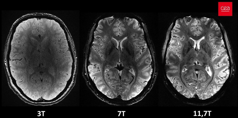
UHF imaging comes with the potential for many new findings and many new challenges. Know your encoding fields to harness the full potential of UHF.
