NeuroCam™ 7T
MR Head Coil for Parallel Transmission with 64 Independent Receive Elements and 16 Field Probes for High Image Quality and Stability at 7T.
- Field fluctuations from system and physiological sources noticeably degrade image quality at ultra-high field strength
- High-performance scanners operating at the technical limit of the hardware inevitably exhibit some system non-linearities that influence image quality
- Non-Cartesian encoding schemes are attractive for advanced neuroimaging but require very accurate knowledge of the encoding fields
- Both, high SNR and spatial resolution are required to answer some of the most fundamental questions about the human brain
MRI has revolutionized our understanding of the human brain by providing non-invasive and detailed insights into its structure and function. The advent of ultra-high field (UHF) MRI scanners, operating at field strengths of 7 Tesla and beyond, has opened up new possibilities for exploring the complexities of the brain. However, along with the numerous advantages offered by UHF imaging, several challenges and considerations must be addressed to ensure optimal image quality and reliable data acquisition. The pursuit of answering fundamental questions about the human brain necessitates both high SNR and spatial resolution in UHF MRI. Achieving these requirements simultaneously presents a technical challenge due to various trade-offs and limitations inherent to UHF systems. Innovations in coil design, parallel imaging techniques, and image reconstruction methods are continuously evolving to strike a balance between SNR and resolution, enabling researchers to obtain the highest quality data for insightful brain investigations. Efforts to mitigate field fluctuations, correct system non-linearities, and optimize non-Cartesian encoding schemes are essential for improving image quality and ensuring reliable data acquisition.
Head Coil with Integrated Field Monitoring for Accurate Neuroimaging at 7 Tesla
NeuroCamTM 7T with its 64 highly sensitive receive elements provides new opportunities for neuroimaging at 7 Tesla. The tight-fitting geometry ensures optimal coil-head coupling, maximizing sensitivity and minimizing signal loss. Additionally, it features an 8 or 16 channel transmit coil, enabling parallel transmission to achieve a homogeneous spin excitation. By elevating SNR and enabling finer spatial resolution, NeuroCamTM 7T captures subtle anatomical details and facilitates the detection of intricate brain activity.
The transmit array and the anterior part of the receiver array can be shifted toward the service end of the magnet for easy subject positioning. The top part of the receiver has indentations on both sides to easily slide it over the subject.

The detachable front-view mirror can be placed on top of the transmit coil to allow the subject to see the magnet room on the patient-side of the bore. The position on the coil can be adjusted via a sliding mechanism.

The tight-fitted receiver array, which comprises 64 independent elements, provides high SNR for neuroimaging applications.

The 8 or 16 independent transmit elements of NeuroCamTM 7T allow to homogenize the transmit field via parallel transmission.

A detachable rear-view mirror can be attached to the enclosure of the receiver array for visual stimulation.

The enclosure of the receiver array is designed to minimally obstruct the visual field.

The compact design and the light weight (14 kg) of the coil assembly facilitates the handling of the coil by the operator.

Susceptibility-Weighted Imaging
Minimum intensity projection
 |
Acquisition parameters
|
T2-Weighted Imaging of the Hippocampus
Acquisition parameters
|
 |
T2 Turbo Spin Echo Imaging
 |
Acquisition parameters
|
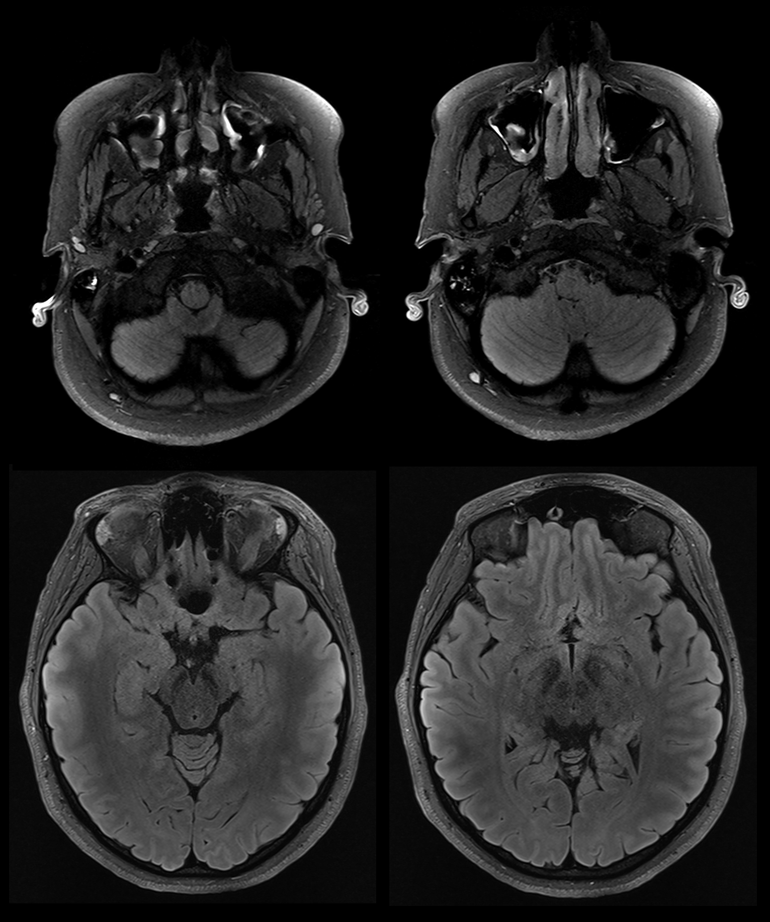
T2 FLAIR FS Turbo Spin Echo Imaging
Acquisition parameters
|
 |
DICOM Viewer
 |
Further Information & Images
NeuroCamTM 7T is available with a fully integrated array of 16 fluorine field probes for concurrent measurement of the encoding fields. The probes are located in the enclosure of the receiver array and distributed around the imaging volume to provide optimal conditioning for the spatial expansion of the spherical harmonics basis fields. The housing of the frontend electronics for the field probe array is attached to the back of the coil, where it does not obstruct the visual field of the rear-view mirror.

Physiological fluctuations
By monitoring the field, adjustments can be made in real time to compensate for fluctuations caused by breathing or other factors, ensuring a more stable and uniform field throughout the imaging procedure. Alternatively, the corrections can be made during image reconstruction by incorporating the measured fields in the process. The figure below shows frequency fluctuations caused by a breathing subject in a 7 T MR scanner as measured by four field probes at different locations in the scanner bore (Courtesy of DZNE Bonn, Germany).

Higher-order field terms
The example below demonstrates the influence of a diffusion preparation on a spiral readout (Courtesy of Sajjad Feizollah and Christine Tardif, McGill University, Montreal, Canada). The data was acquired with a Clip-on Camera on a 7T scanner. The animation on the left-hand side illustrates a field-monitored spiral trajectory that was not preceded by a diffusion gradient. Upon closer examination, distinct characteristics differentiate it from its counterpart on the right-hand side that is influenced by a preceding diffusion gradient. Particularly noteworthy is the considerable disparity observed in the zeroth order term (k0), which represents the fluctuation of the global magnetic field. Upon magnification, discernible amplification of higher order fields is observed beyond the zeroth term. These amplified higher-order fields can be attributed to eddy currents induced by the diffusion gradients. Higher-order terms can have a direct impact on image quality and geometric consistency, which makes their characterization very important.
 |
 |
The ability to simultaneously measure the actual encoding fields provides the opportunity to select more demanding yet efficient readout schemes. To illustrate this, intra-cellular volume fraction (ICVF) maps derived from diffusion-weighted data acquired using two different readout techniques are shown below: echo planar imaging (EPI) on the left and spiral imaging on the right. The usage of a spiral readout, which achieves higher SNR, directly translates into ICVF maps of superior quality.
By default, NeuroCamTM 7T is equipped with eight independent transmit and 64 receive elements. It includes both, a front and a rear-view mirror for patient comfort and visual stimulation.

NeuroCamTM 7T is also available with 16 independent transmit channels for parallel excitation. The coil does not comprise a front-view mirror due to the smaller size of the transmit elements, but it includes the same rear-view mirror for visual stimulation as the 8-channel version of the coil.

The coil frame can be adapted to fit into a head-only gradient system if the latter is large enough to accommodate the transmit coil. Note that a version with an integrated field probe array for head-only gradient systems is currently not available for purchase but might be in the future.

Versions
NeuroCamTM 7T exists in three versions, each tailored to meet specific imaging needs. Choose from our Standard, Advanced, and Excellence versions, each offering unique features and capabilities for exceptional neuroimaging performance.
 |
This product is developed in collaboration with MR CoilTech. |
Some features of the product are still under development and not commercially available yet. Their future availability cannot be ensured.All data and information contained on this webpage are legally not binding and shall not create any warranties or liabilities whatsoever of Skope.CAUTION – Investigational device. Limited by Federal law to investigational use.
|